
What is LLaVA-Rad
LLaVA-Rad is a small multimodal model launched by Microsoft Research, focused on clinical radiology report generation. It is a branch of the LLaVA-Med project, specifically for chest X-ray (CXR) imaging. Based on LLaVA-Med's foundation architecture and training methods, it has been optimized for the specific needs of the radiology field. Through modular training, combining unimodal pretraining, alignment, and fine-tuning in three stages, it embeds non-text modalities such as images into the text space based on an adapter mechanism, achieving efficient training and inference. The model is trained on 697,435 pairs of radiology images and reports, with excellent performance; key metrics such as ROUGE and -LF1-RadGraph improved by 12.1% and 10.1%, respectively. LLaVA-Rad is designed to be lightweight, requiring only a single V100 GPU to run, and training can be completed within one day, making it suitable for rapid clinical deployment. A companion CheXprompt automatic evaluation metric was also launched, used to score the factual correctness of reports.
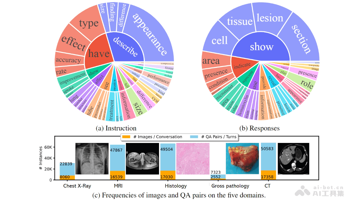
Main features of LLaVA-Rad
- Radiology report generation: The core function of LLaVA-Rad is to automatically generate high-quality radiology reports, especially for chest X-ray (CXR) imaging. It can generate detailed diagnostic reports based on input medical images, helping doctors quickly and accurately record and communicate examination results.
- Multimodal fusion: Through a modular training method, the model embeds non-text modalities such as images into the text embedding space. Based on an efficient adapter mechanism, it achieves effective fusion of images and text, generating more accurate reports.
- Efficient training and inference: LLaVA-Rad is designed to be lightweight, requiring only a single V100 GPU to complete inference, and training can be completed within one day.
- Automatic evaluation and quality control: To better evaluate the factual correctness of reports, LLaVA-Rad has launched CheXprompt, an automatic scoring metric based on GPT-4. It can effectively solve evaluation challenges in clinical applications and ensure that generated reports meet medical standards.
Technical principles of LLaVA-Rad
- Modular training method: The training process of LLaVA-Rad is divided into three stages: unimodal pretraining, alignment, and fine-tuning.
- Unimodal pretraining: First, text and images are pretrained separately to learn their respective feature representations.
- Alignment: Through an adapter mechanism, image features are embedded into the text embedding space, achieving alignment between images and text.
- Fine-tuning: Fine-tuning is performed on the aligned multimodal data to further optimize model performance.
- Performance optimization
- Lightweight design: LLaVA-Rad is a small multimodal model that requires only one V100 GPU to complete inference, and training can be completed within one day.
- Diverse datasets: The model is trained on a dataset containing 697,435 pairs of radiology images and reports, with data from seven different sources, ensuring the model's generalization ability.
- Performance improvement: On key metrics (such as ROUGE-L and F1-RadGraph), LLaVA-Rad improved by 12.1% and 10.1%, respectively, compared with other similar models.
Project address of LLaVA-Rad
- Github repository:https://github.com/microsoft/LLaVA-Med
- arXiv technical paper:https://arxiv.org/pdf/2306.00890
Application scenarios of LLaVA-Rad
- Automatic radiology report generation: LLaVA-Rad can automatically generate high-quality radiology reports, helping radiologists quickly and accurately record examination results.
- Clinical Decision Support: By generating detailed radiology reports, LLaVA-Rad provides important decision support for clinicians, especially when dealing with complex diseases, helping doctors quickly identify key findings and make diagnoses.
- Medical Image Analysis: LLaVA-Rad focuses on chest X-ray imaging, enabling rapid analysis of medical images and generation of corresponding reports.