
What is EchoCare
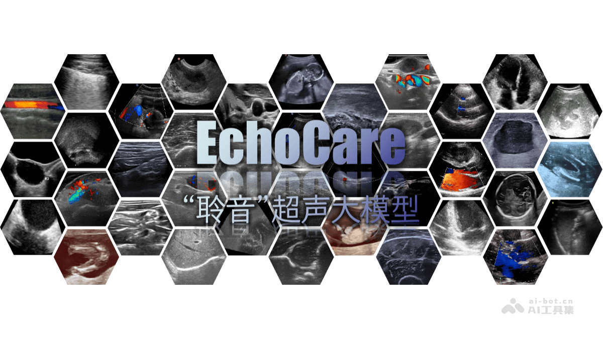
EchoCare is an ultrasound large model launched by the Center for Artificial Intelligence and Robotics (CAIR) of the Hong Kong Institute of Science & Innovation, Chinese Academy of Sciences. The model is trained on the EchoAtlas dataset of 4.5 million ultrasound images, which covers images collected from 23 clinical centers across 5 continents and 38 imaging devices, covering 9 major regions of the human body and 52 anatomical organs, making it one of the largest ultrasound image datasets currently available. EchoCare adopts a "structured contrastive self-supervised learning framework" and, through techniques such as image mask reconstruction and adaptive hard patch mining, significantly improves the model's ability to model the deep semantics of ultrasound images and its generalization performance. In seven major medical tasks, including ultrasound image segmentation, classification, detection, regression, and enhancement, its performance surpasses current state-of-the-art methods.
Main Functions of EchoCare
-
Ultrasound Image Segmentation: It can precisely segment different tissues and organs in ultrasound images, helping doctors more clearly identify the boundaries between lesion areas and normal tissues, and providing a more accurate basis for diagnosis.
-
Lesion Classification: It classifies lesions in ultrasound images, such as distinguishing benign from malignant tumors, assisting doctors in quickly determining the nature of lesions and improving diagnostic efficiency.
-
Organ Detection and Segmentation: It can not only detect the positions of organs in images, but also precisely segment organs, providing detailed anatomical information for subsequent diagnosis and treatment.
-
Image Enhancement: It improves the quality of ultrasound images, enhancing image contrast and clarity, enabling doctors to more clearly observe subtle structures and lesions and reducing the possibility of misdiagnosis.
-
Report Generation: It automatically generates diagnostic reports based on ultrasound image analysis results, saving doctors time in writing reports, improving work efficiency, and ensuring the accuracy and consistency of reports.
Technical Principles of EchoCare
-
Large-Scale Dataset Construction: It is trained on the EchoAtlas dataset of 4.5 million ultrasound images. The dataset covers a global cohort spanning multiple centers, multiple devices, and multiple ethnic groups, covering 9 major regions of the human body and 52 anatomical organs, providing rich learning material for the model.
-
Structured Contrastive Self-Supervised Learning Framework: It introduces hierarchical tree-structured labels based on medical priors, enabling structured learning and implicit encoding of multi-label semantic relationships, and improving the model's ability to model the deep semantics of ultrasound images.
-
Image Mask Reconstruction Technology: Through mask reconstruction of images, it enhances the model's learning and understanding of local image features, improving the model's robustness and generalization performance.
-
Adaptive Hard Patch Mining Technology: It automatically identifies and focuses on image regions that are difficult to learn, strengthens learning in a targeted manner, and improves the model's ability to handle complex images.
-
Progressive Training Strategy: It adopts a progressive training method, gradually increasing the difficulty and complexity of model training, helping the model better adapt to different types of ultrasound images and improving overall performance.
EchoCare Project Address
- Project Official Website:https://echocare.cares-copilot.com/
- Github Repository:https://github.com/CAIR-HKISI/EchoCare
- arXiv Technical Paper:https://arxiv.org/pdf/2509.11752
Application Scenarios of EchoCare
-
Routine Hospital Examinations: Applied in routine ultrasound examinations in hospitals, it can significantly reduce dependence on professionals, assist doctors in diagnosing more efficiently and accurately, effectively improve the efficiency of medical services, and provide more possibilities for the optimal allocation of medical resources.
-
Disease Diagnosis and Screening: It can be used for the diagnosis and screening of various diseases, as verified by specific cases in 1,556 ultrasound cases of ovarian tumors in the Department of Obstetrics and Gynecology at Qilu Hospital of Shandong University and more than 1,000 thyroid ultrasound examinations at Xiangya Hospital of Central South University, and its performance is significantly superior to existing SOTA methods.
-
Cardiac Ultrasound Detection: In the detection and analysis of cardiac ultrasound aortic aneurysms, retrospective validation can be conducted, providing strong support for the diagnosis of cardiac diseases.
-
Ultrasound Image Processing: It includes seven major medical tasks such as ultrasound image segmentation, classification, detection, regression, and enhancement, as well as more than ten downstream applications, such as lesion classification, organ detection and segmentation, and image enhancement, improving the quality and diagnostic value of ultrasound images.
-
Clinical Adaptation and Research: The foundation model is planned to be open-sourced for use by medical institutions, and subsequently prospective research, emergency room scenario applications, and hardware integration jointly with ultrasound device companies will be advanced in stages.